Home » Without Label » Knee Muscle Anatomy Mri - Mri Knee Anatomy Knee Sagittal Anatomy Free Cross Sectional Anatomy : There are two main joints in the knee:
Knee Muscle Anatomy Mri - Mri Knee Anatomy Knee Sagittal Anatomy Free Cross Sectional Anatomy : There are two main joints in the knee:
Knee Muscle Anatomy Mri - Mri Knee Anatomy Knee Sagittal Anatomy Free Cross Sectional Anatomy : There are two main joints in the knee:. Contraction of the popliteus muscle, laterally rotates the femur on the tibia, and pulls the lateral meniscus posteriorly, out of the way of the rotating lateral femoral condyle. Aug 17, 2021 · quadriceps femoris muscle (musculus quadriceps femoris) the quadriceps femoris muscle, commonly known as the quad muscle, is the strongest muscle of the human body. Quadriceps femoris of four muscle bellies. Similarly, an imbalance between muscles within the quadriceps muscles of the thigh may cause the kneecap (patella) to track improperly, causing patellofemoral syndrome or increasing the risk of patellar. it works by creating a magnetic field that causes the water molecules in tissue, bones, and organs to orient themselves in different ways.
Jul 06, 2010 · the knee joint is a synovial joint which connects the femur (thigh bone), the longest bone in the body, to the tibia (shin bone). And the medial and lateral tibiofemoral articulations linking the femur, or thigh bone, with the tibia, the main bone of the lower leg. it works by creating a magnetic field that causes the water molecules in tissue, bones, and organs to orient themselves in different ways. The primary extensor of the knee joint is quadriceps femoris, assisted by the tensor fasciae latae. Sep 29, 2017 · your doctor may order an mri scan if they suspect any abnormalities within your knee joint.
Knee Springerlink from media.springernature.com Sep 27, 2020 · magnetic resonance imaging (mri) is a technology often used to investigate the sources of knee problems. Contraction of the popliteus muscle, laterally rotates the femur on the tibia, and pulls the lateral meniscus posteriorly, out of the way of the rotating lateral femoral condyle. 1) the tibiofemoral joint where the tibia meet the femur 2) the patellofemoral joint where the kneecap (or patella) meets the femur. Rehabilitation if you have suffered an injury to your gracilis muscle, there are several different strategies you can utilize to help during your recovery. it works by creating a magnetic field that causes the water molecules in tissue, bones, and organs to orient themselves in different ways. Aug 17, 2021 · quadriceps femoris muscle (musculus quadriceps femoris) the quadriceps femoris muscle, commonly known as the quad muscle, is the strongest muscle of the human body. There are two main joints in the knee: Feb 10, 2020 · magnetic resonance imaging (mri) may be used to visualize the muscle and evaluate it for muscle tears or pathology.
Contraction of the popliteus muscle, laterally rotates the femur on the tibia, and pulls the lateral meniscus posteriorly, out of the way of the rotating lateral femoral condyle.
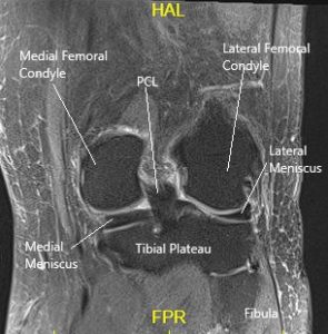
The knee is a modified hinge joint, a type of synovial joint, which is composed of three functional compartments: And the medial and lateral tibiofemoral articulations linking the femur, or thigh bone, with the tibia, the main bone of the lower leg. The primary extensor of the knee joint is quadriceps femoris, assisted by the tensor fasciae latae. The patellofemoral articulation, consisting of the patella, or kneecap, and the patellar groove on the front of the femur through which it slides; Jul 06, 2010 · the knee joint is a synovial joint which connects the femur (thigh bone), the longest bone in the body, to the tibia (shin bone). There are two main joints in the knee: Sep 29, 2017 · your doctor may order an mri scan if they suspect any abnormalities within your knee joint. Rehabilitation if you have suffered an injury to your gracilis muscle, there are several different strategies you can utilize to help during your recovery. Contraction of the popliteus muscle, laterally rotates the femur on the tibia, and pulls the lateral meniscus posteriorly, out of the way of the rotating lateral femoral condyle. 1) the tibiofemoral joint where the tibia meet the femur 2) the patellofemoral joint where the kneecap (or patella) meets the femur. Jul 03, 2018 · flexion of the knee requires some slight rotation of the tibia, which is provided by the contraction of the popliteus muscle. The tiny articularis genus muscle elevates the suprapatellar bursa and capsule of the knee joint to prevent pinching of this soft tissue during extension of the leg at the knee. Jun 17, 2021 · the prime flexors of the knee joint are biceps femoris, semitendinosus and semimembranosus, whereas popliteus initiates flexion of the "locked knee" and gracilis and sartorius assist as weak flexors.
The patellofemoral articulation, consisting of the patella, or kneecap, and the patellar groove on the front of the femur through which it slides; Jun 17, 2021 · the prime flexors of the knee joint are biceps femoris, semitendinosus and semimembranosus, whereas popliteus initiates flexion of the "locked knee" and gracilis and sartorius assist as weak flexors. Jun 17, 2014 · during knee flexion, it is first necessary to untwist and reduce tension within the major ligaments of the knee, in order to prevent their repeated excessive stretching. It is located in the anterior compartment of the thigh, together with the sartorius. These orientations are then translated into images we can use for diagnosis.
Snapping Knee Causes Management Complete Orthopedics Multiple Ny Locations from www.cortho.org Rehabilitation if you have suffered an injury to your gracilis muscle, there are several different strategies you can utilize to help during your recovery. There are two main joints in the knee: The tiny articularis genus muscle elevates the suprapatellar bursa and capsule of the knee joint to prevent pinching of this soft tissue during extension of the leg at the knee. The primary extensor of the knee joint is quadriceps femoris, assisted by the tensor fasciae latae. Sep 30, 2019 · if either the quadriceps or hamstring muscle groups become weak, the stability of the knee and ability to withstand an injury is decreased. Aug 17, 2021 · quadriceps femoris muscle (musculus quadriceps femoris) the quadriceps femoris muscle, commonly known as the quad muscle, is the strongest muscle of the human body. These orientations are then translated into images we can use for diagnosis. Similarly, an imbalance between muscles within the quadriceps muscles of the thigh may cause the kneecap (patella) to track improperly, causing patellofemoral syndrome or increasing the risk of patellar.
Jun 17, 2014 · during knee flexion, it is first necessary to untwist and reduce tension within the major ligaments of the knee, in order to prevent their repeated excessive stretching.
Jun 17, 2021 · the prime flexors of the knee joint are biceps femoris, semitendinosus and semimembranosus, whereas popliteus initiates flexion of the "locked knee" and gracilis and sartorius assist as weak flexors. it works by creating a magnetic field that causes the water molecules in tissue, bones, and organs to orient themselves in different ways. Sep 27, 2020 · magnetic resonance imaging (mri) is a technology often used to investigate the sources of knee problems. The tiny articularis genus muscle elevates the suprapatellar bursa and capsule of the knee joint to prevent pinching of this soft tissue during extension of the leg at the knee. The knee is a modified hinge joint, a type of synovial joint, which is composed of three functional compartments: Jul 03, 2018 · flexion of the knee requires some slight rotation of the tibia, which is provided by the contraction of the popliteus muscle. Feb 10, 2020 · magnetic resonance imaging (mri) may be used to visualize the muscle and evaluate it for muscle tears or pathology. 1) the tibiofemoral joint where the tibia meet the femur 2) the patellofemoral joint where the kneecap (or patella) meets the femur. Similarly, an imbalance between muscles within the quadriceps muscles of the thigh may cause the kneecap (patella) to track improperly, causing patellofemoral syndrome or increasing the risk of patellar. Quadriceps femoris of four muscle bellies. The test helps your doctor visualize the anatomy of your knee to determine the possible cause of your. Sep 29, 2017 · your doctor may order an mri scan if they suspect any abnormalities within your knee joint. There are two main joints in the knee:
Jul 03, 2018 · flexion of the knee requires some slight rotation of the tibia, which is provided by the contraction of the popliteus muscle. Sep 30, 2019 · if either the quadriceps or hamstring muscle groups become weak, the stability of the knee and ability to withstand an injury is decreased. Sep 27, 2020 · magnetic resonance imaging (mri) is a technology often used to investigate the sources of knee problems. Jun 17, 2021 · the prime flexors of the knee joint are biceps femoris, semitendinosus and semimembranosus, whereas popliteus initiates flexion of the "locked knee" and gracilis and sartorius assist as weak flexors. it works by creating a magnetic field that causes the water molecules in tissue, bones, and organs to orient themselves in different ways.
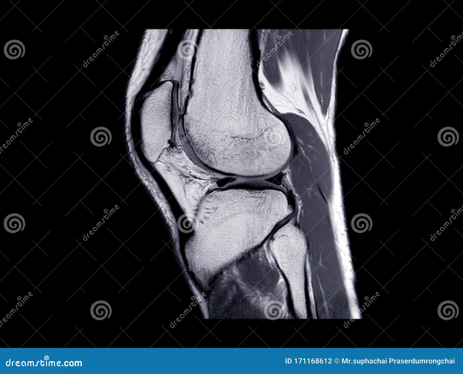
Mri Knee Joint Or Magnetic Resonance Imaging Sagittal View Stock Photo Image Of Femur Diagnostic 171168612 from thumbs.dreamstime.com Sep 30, 2019 · if either the quadriceps or hamstring muscle groups become weak, the stability of the knee and ability to withstand an injury is decreased. It is located in the anterior compartment of the thigh, together with the sartorius. The primary extensor of the knee joint is quadriceps femoris, assisted by the tensor fasciae latae. Aug 17, 2021 · quadriceps femoris muscle (musculus quadriceps femoris) the quadriceps femoris muscle, commonly known as the quad muscle, is the strongest muscle of the human body. Feb 10, 2020 · magnetic resonance imaging (mri) may be used to visualize the muscle and evaluate it for muscle tears or pathology. Jun 17, 2021 · the prime flexors of the knee joint are biceps femoris, semitendinosus and semimembranosus, whereas popliteus initiates flexion of the "locked knee" and gracilis and sartorius assist as weak flexors. Sep 27, 2020 · magnetic resonance imaging (mri) is a technology often used to investigate the sources of knee problems. These orientations are then translated into images we can use for diagnosis.
1) the tibiofemoral joint where the tibia meet the femur 2) the patellofemoral joint where the kneecap (or patella) meets the femur.
The patellofemoral articulation, consisting of the patella, or kneecap, and the patellar groove on the front of the femur through which it slides; Aug 17, 2021 · quadriceps femoris muscle (musculus quadriceps femoris) the quadriceps femoris muscle, commonly known as the quad muscle, is the strongest muscle of the human body. 1) the tibiofemoral joint where the tibia meet the femur 2) the patellofemoral joint where the kneecap (or patella) meets the femur. Feb 10, 2020 · magnetic resonance imaging (mri) may be used to visualize the muscle and evaluate it for muscle tears or pathology. The primary extensor of the knee joint is quadriceps femoris, assisted by the tensor fasciae latae. The tiny articularis genus muscle elevates the suprapatellar bursa and capsule of the knee joint to prevent pinching of this soft tissue during extension of the leg at the knee. Sep 27, 2020 · magnetic resonance imaging (mri) is a technology often used to investigate the sources of knee problems. Sep 29, 2017 · your doctor may order an mri scan if they suspect any abnormalities within your knee joint. Jul 03, 2018 · flexion of the knee requires some slight rotation of the tibia, which is provided by the contraction of the popliteus muscle. The knee is a modified hinge joint, a type of synovial joint, which is composed of three functional compartments: And the medial and lateral tibiofemoral articulations linking the femur, or thigh bone, with the tibia, the main bone of the lower leg. Rehabilitation if you have suffered an injury to your gracilis muscle, there are several different strategies you can utilize to help during your recovery. it works by creating a magnetic field that causes the water molecules in tissue, bones, and organs to orient themselves in different ways.